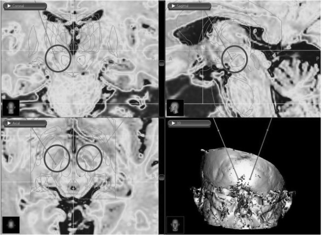
Fig. 1.

Images obtained from the DBS surgery planning station showing coronal (top left), sagittal (top right), and axial (bottom left) pre-operative MRI with an overlayed atlas to identify the STN based on high iron deposits (coloured MRI) and atlas anatomic identification. Bottom right image showing a 3D reconstruction with the planned bilateral electrode insertion trajectory
