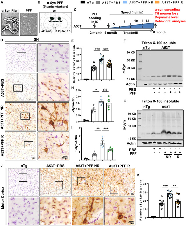
Figure 1. Treadmill inhibits α-syn spreading in A53T mice brain.
(A) Preformed α-syn fibril (PFF) that was validated by electron microscopy.
(B and C) PFF was bilaterally injected into the internal capsule (IC) of 2-month-old A53T mice (B), and following 2 months of PFF injection, animals were run in a treadmill for the next 2 months (C).
(D–I) Spreading of α-syn in brains of PFF-seeded non-running (A53T + PFF NR) and running (A53T + PFF R) mice was evaluated by phosphoserine 129 (pSyn129) staining in midbrain sections (F3,36 = 166.2, p < 0.001, D and E) and immunoblotting of total Triton X-100 soluble and insoluble α-syn in the SN (F3,16 = 32.18, p < 0.01, F–I).
(J and K) Propagation of α-syn to motor cortex was evaluated by pSyn129 immunostaining followed by quantification (F3, 36 = 102.4, p < 0.01).
For immunostaining, at least two sections from each brain per group were stained, and 10–15 pSyn129-positive cells were quantified from each section by Fiji software. Data were analyzed by one-way ANOVA followed by Tukey’s multiple comparison tests. *p < 0.05, **p < 0.01, ***p < 0.001 indicate significance compared with respective groups; ns, not significant. Data are represented as mean ± SEM (n = 5 animals per each group).

