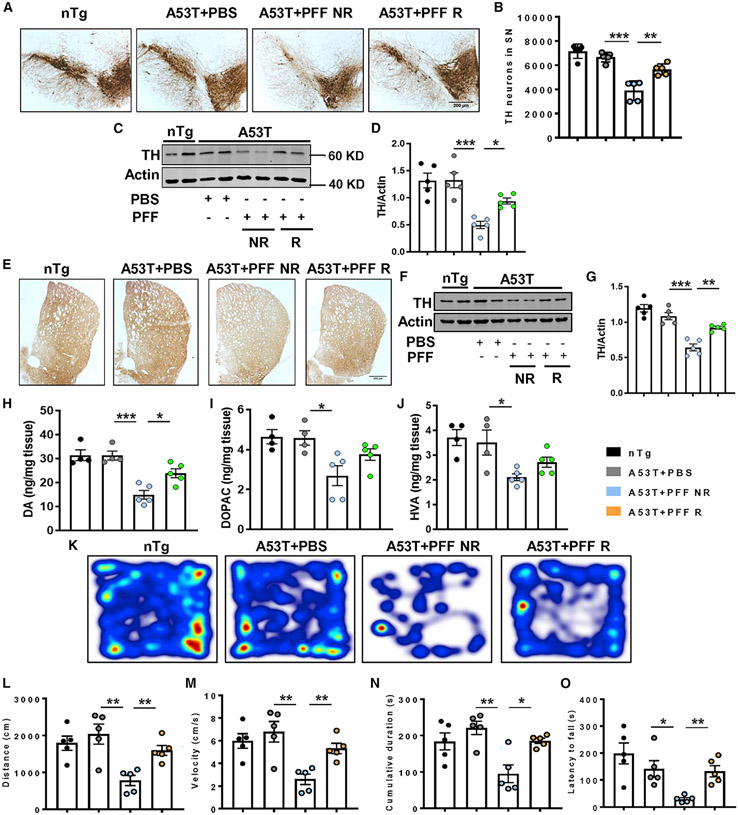
Figure 2. Attenuation of parkinsonian features by treadmill in PFF-seeded A53T mice.
(A–G) PFF-induced parkinsonian pathology was determined by tyrosine hydroxylase (TH) staining in nigral sections (A), stereological counting of TH-positive neurons (F3, 16 = 29.97, p < 0.01, B), total TH level in SN (F3, 16 = 13.67, p < 0.05, C and D), TH fiber staining (E), and total TH level (F3, 16 = 28.37, p < 0.01, F and G) in striatum of different groups of animals.
(H–J) Dopamine (DA) and its metabolites, 3,4-dihyroxyphenyl acetic acid (DOPAC) and homovanillic acid (HVA), were measured from striatal tissues by high-performance liquid chromatography electrochemical detection (HPLC-ECD; F3, 14 = 16.18, p < 0.05 for DA).
(M–Q) Behavioral deficit in PFF-seeded animals was monitored by open-field locomotor test (F3, 16 = 8.04, p < 0.01 for distance, F3, 16 = 8.04, p < 0.01 for velocity, and F3, 16 = 7.602, p < 0.05 for cumulative duration, M–P) and rotarod test (F3, 16 = 14.49, p < 0.01, Q).
Data were statistically analyzed by unpaired two-tailed t test for comparison between two samples and by one-way ANOVA followed by Tukey’s multiple comparison tests. *p < 0.05, **p < 0.01, ***p < 0.001, ns, not significant. Values are represented as mean ± SEM (n = 5 animals per each group).

