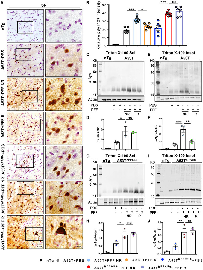
Figure 6. Treadmill exercise does not reduce α-syn spreading in the brain in the absence of PPARα.
Age-matched A53T and A53T mice lacking functional PPARα (A53TΔPPARα) were injected with either PBS or PFF at the IC region of striatum, and following 2 months of seeding, animals were run in a treadmill daily for 2 months.
(A) IHC of pSyn129 in the SN followed by relative pSyn129 intensity analysis.For immunostaining, at least two sections from each brain per group were stained, and 10–15 pSyn129-positive cells were quantified in each section.
(B) Average intensity of pSyn129 obtained from each section of all groups of mice are shown (F6, 35 = 62.92, *p < 0.05, ***p < 0.001, n = 3).
(C, E, G, and I) The levels of total α-syn in the Triton X-100 soluble and insoluble fractions isolated from midbrain tissues (C and E) and A53TΔPPARα mice (G and I) were measured by immunoblotting.
(D, F, H, and J) Ratio of band intensities of α-syn to actin (D and H for soluble fractions and F and J for insoluble fractions, F4, 11 = 24.85, **p < 0.01 for A53T + PFF NR versus A53T + PFF R for insoluble fraction, n = 3).
The significance of mean was compared using one-way ANOVA followed by Tukey’s multiple comparison test. *p < 0.05, **p < 0.01, and ***p < 0.001; ns, not significant. Data are represented as mean ± SEM.

