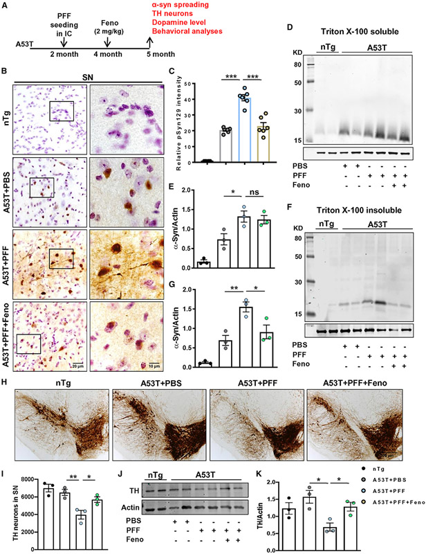
Figure 7. Oral fenofibrate inhibits α-syn spreading and protects nigral DAergic neurons in PFF-seeded A53T mice brain.
(A) PFF-seeded A53T mice were fed with fenofibrate (2 mg/kg) daily via gavage for 1 month.
(B) Spreading of α-syn in SN was analyzed by pSyn129 IHC.
(C) Relative intensity of pSyn129 (F3, 20 = 109.7, ***p < 0.001, n = 3).
(D–G) The level of α-syn in Triton X-100 soluble (D and E) and insoluble tissue fractions (F3, 11 = 21.38 for A53T + PFF versus A53T + PFF + Feno, *p < 0.05, **p < 0.01, F and G, n = 3) was assessed by immunoblotting.
(H–K) Number of TH neurons in SN was shown by IHC of midbrain sections (H) followed by stereological counting in each hemisphere of mouse brain (*p < 0.05, **p < 0.01, I, n = 3), and the TH protein content in SN was measured by immunoblotting (*p < 0.05, J and K, n = 3).
The significance of mean was compared using one-way ANOVA followed by Tukey’s multiple comparison test. *p < 0.05, **p < 0.01, and ***p < 0.001 indicate significance compared to respective groups; ns, not significant. Data are represented as mean ± SEM.

