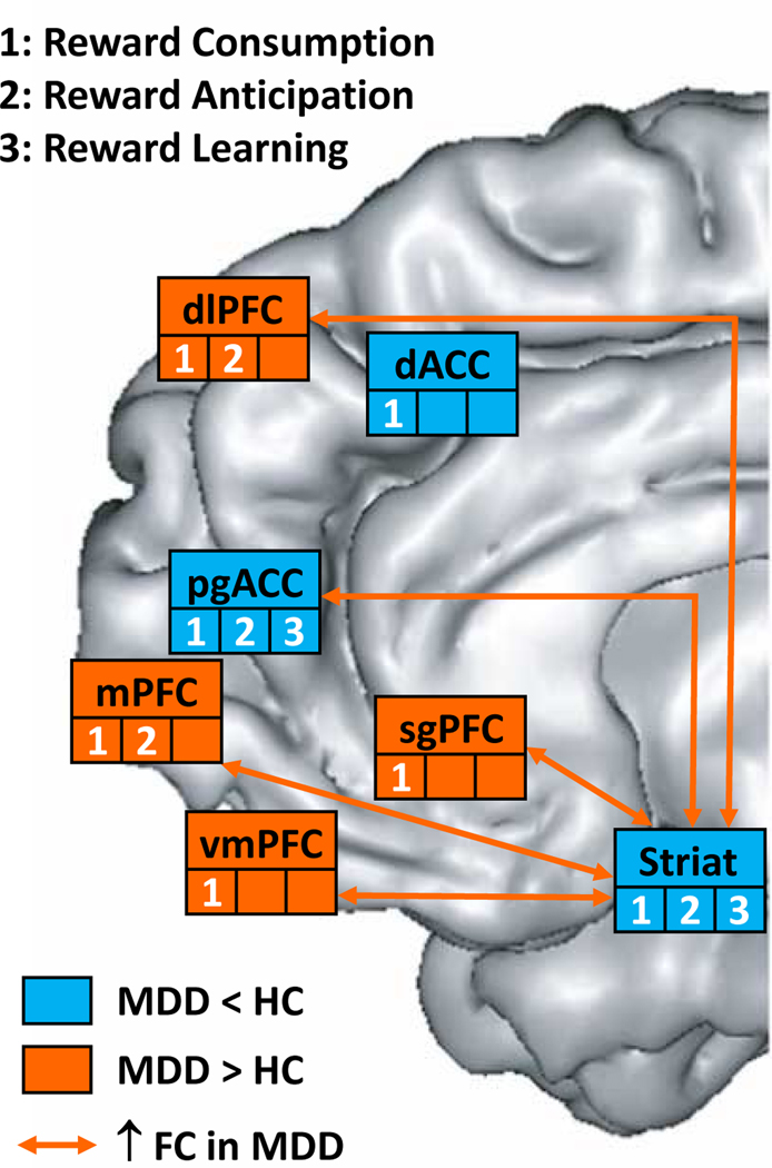
Figure 2:
Summary of abnormalities emerging from functional magnetic resonance imaging (fMRI) in individuals with major depressive disorder (MDD) or at risk for MDD using tasks probing reward-related processes or evaluating resting state functional connectivity within the brain reward system. Regions highlighted in orange and blue show higher activation and lower activation, respectively, in MDD samples than healthy controls (HC). Orange arrows denote higher functional connectivity in MDD samples than healthy controls. dACC: dorsal anterior cingulate cortex, dlPFC: dorsolateral prefrontal cortex, mPFC: medial prefrontal cortex, pgACC; perigenual anterior cingulate cortex, sgACC: subgenual anterior cingulate cortex, Striat: striatum vmPFC: ventromedial prefrontal cortex. Modified after (34).

