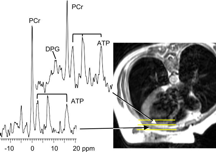
Figure 1. Representative CMR image and 31P MR spectra.
Representative axial MRI (right) of the chest of a 45-year-old woman (lying prone) who subsequently experienced an arrhythmic event. Yellow lines denote localized volumes from which 31P MRS spectra (left) were derived (arrows). The upper spectrum from the heart shows phosphocreatine (PCr), diphosphoglycerate (DPG), and the 3 phosphate resonances of ATP. The myocardial ATP was low (2.5 μmol/g wet wt) but PCr/ATP was normal (1.7) in this individual. The lower spectrum includes chest muscle and is shown for comparison but was not used in analysis.

