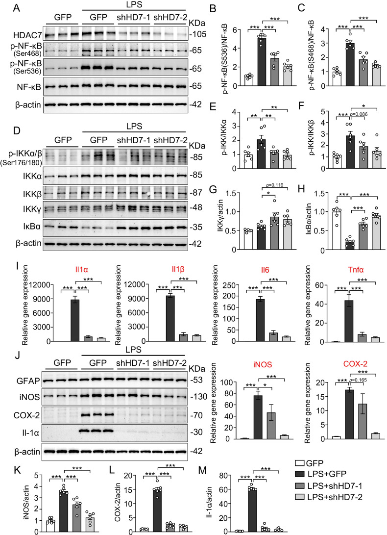
Fig. 4.
Genetic knockdown of HDAC7 suppresses LPS-induced NF-κB activation and inflammatory responses in primary cultured astrocytes. A–C Knockdown of HDAC7 inhibits LPS-induced NF-κB activation in cultured astrocytes. A Representative western blots showing protein levels of HDAC7, p-NF-κB(S468), p-NF-κB(S536), NF-κB, and β-actin in control and LPS-treated primary cultured mouse astrocytes (astrocytes were infected with Lenti-GfaABC1D-HDAC7-T2A-GFP or Lenti-GfaABC1D-T2A-GFP for 5 days, and then treated with LPS (100 ng/ml) for 24 h). B Quantification analysis of p-NF-κB(S468) normalized to total NF-κB. C Quantification analysis of p-NF-κB(S536) normalized to total NF-κB. N = 6 for each group, one-way ANOVA, Dunnett’s post hoc analysis. D–H Knockdown of HDAC7 suppresses IκBα degradation with little effects on IKK expression. D Representative western blots showing protein levels of p-IKKα/β(S176/180), IKKα, IKKβ, IKKγ, IκBα, and β-actin in control and LPS-stimulated primary mouse astrocytes as described above. Quantification of p-IKKα/β(S176/180) normalized to IKKα (E), p-IKKα/β(S176/180) normalized to IKKβ (F), IKKγ normalized to β-actin (G), and IκBα normalized to β-actin (H). N = 6 for each group, one-way ANOVA, Dunnett’s post hoc analysis. I–M Knockdown of HDAC7 inhibits LPS-induced inflammatory responses in cultured astrocytes. I RT-qPCR analysis of Il-1α, Il-1β, Il6, Tnfα, iNOS, and COX-2 in control and LPS-stimulated primary cultured mouse astrocytes as described above. J Representative western blots showing protein levels of GFAP, iNOS, COX-2, Il-1α, and β-actin in control and LPS-treated primary cultured mouse astrocytes. Quantification of iNOS (K), COX-2 (L), and Il-1α (M) normalized to β-actin. N = 6 for each group, one-way ANOVA, Dunnett’s post hoc analysis. Data were expressed as mean ± SEM, *p < 0.05, **p < 0.01, ***p < 0.001

