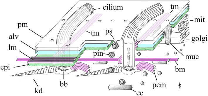
FIGURE 4.

Organization of the cell cortex in Tetrahymena. A patch of cell cortex is depicted with anterior to the left, and posterior to the right. pm = plasma membrane; alv = Ca++ reservoirs known as alveolar sacs; lm = longitudinal band of microtubules lying just under the alveolar sacs and above the epiplasm; epi = a proteinaceous layer of nonmicrotubule based cytoskeleton resembling the spectrin layer in red blood cells; tm = transverse microtubules exposed to cytoplasm; kd = the kinetodesma (or striated fiber); bb = BB; pin = pinosomes; ps = parasomal sac, site of clathrin‐mediated pinocytosis; ee = early endosomes; pcm = postciliary microtubules (also exposed to cytoplasm) and the bm = basal microtubule: the one microtubule track that runs the length of the cell (anterior to posterior) that is exposed to the cytoplasm and hence available for A/P vesicle traffic; muc = mucocyst (dense‐core secretory granule); golgi = dictyosome; mit = mitochondrion
