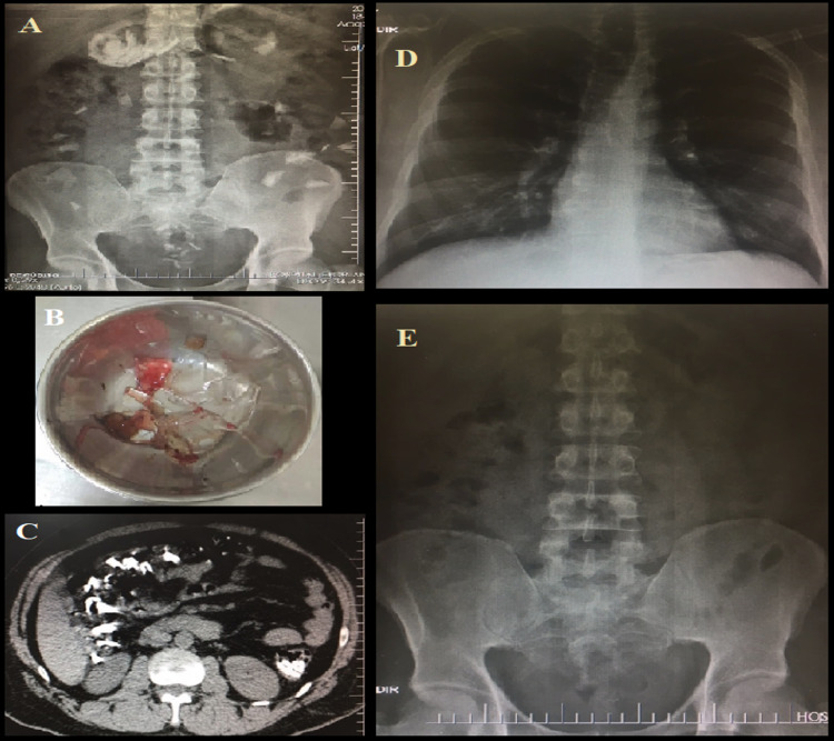
Figure 1. Radiographic and upper gastrointestinal endoscopy findings.
Panel A: radiograph of an orthostatic abdomen (first hospitalization day) showing several radiopaque fragments (glass) inside the stomach and in the intestine.
Panel B: metal container holding several glass shards that were removed from the patient's stomach through upper gastrointestinal endoscopy.
Panel C: total abdomen tomography (without intravenous contrast) performed for control purposes (second hospitalization day) showing glass shards inside the intestine, although with no signs of pneumoperitoneum.
Panels D-E: radiological control performed on the last hospitalization day showing the complete elimination of the glass shards and lack of pathological intracavitary air signs.

