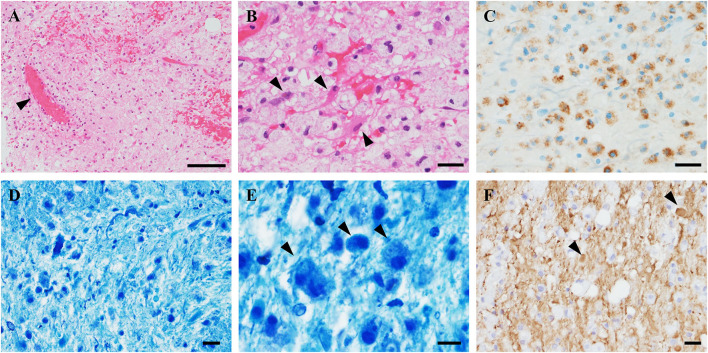
Figure 2.
Pathological findings of the brain biopsy. (A) Hematoxylin and eosin (HE) stains showed gliosis with widespread infiltration of inflammatory cells without neoplastic tissue. Perivascular inflammation was absent (arrowhead). (B) High-magnification HE showed diffuse foamy macrophage infiltration associated with reactive astrocytes (arrowheads). (C) CD68-immunostaining revealed clusters of macrophages. (D,E) Klüver–Barrera staining demonstrated myelin loss with myelin-laden macrophages (arrowheads). (F) Phosphorylated neurofilament immunostaining revealed axonal fragmentation and spheroids (arrowheads). The axonal loss was milder than myelin loss. Scale bars: (A) 100 μm. (B–E) 20 μm. (F) 10 μm.

