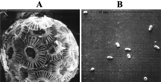
FIG. 1.
Scanning electron micrographs of E. huxleyi strain 1516 life cycle types. (A) Nonmotile C cell showing overlapping coccolith plates. (B) Motile S cells representing a possible gametic stage of the life cycle. Note the relative difference in the sizes of these two cell types involved in phase variation mechanisms described in the text. Magnifications, ca. ×8,000 (A) and ×10,500 (B).

