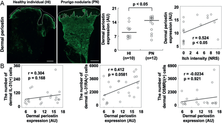
Fig. 1.
Periostin in prurigo nodularis (PN). Representative images of PN lesions and healthy individual (HI) skin with quantification of staining. (A) Dermal expression of periostin was significantly enhanced in PN lesions and correlated with itch intensity. White dotted circles indicate the epidermis excluding the stratum corneum. Black lines in the center panel represent medians. (B) Dermal periostin did not correlate with the number of dermal IL- 31(+) cells, IL-31RA(+) cells, and OSMRβ(+) cells. Bar = 1 mm. AU: arbitrary unit; r: Spearman’s rank correlation coefficient; NRS: numerical rating scale.

