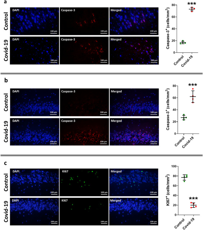
Fig. 6.
Immunofluorescence staining of cleaved caspases-3 in tissue sections taken from the hippocampal pyramidal layer (a) and granular layer (b) of dentate gyrus, and neurogenesis factor Ki67 (c) in dentate gyrus of two groups of cases including control and COVID-19. The antibody in detecting cleaved caspases-3, and Ki67 are shown in right side (red is cleaved caspase-3 staining, green is KI67, and total nuclei stained with DAPI are blue) (a and b), and the left graphs illustrate in the mean and standard error of cleaved caspase-3 and Ki67 marker expression in dentate gyrus. Asterisk (*) shows the difference between the COVID and the control group (*P < 0.05; **P < 0.01; ***P < 0.001). The values were expressed as means ± SEM. The values were expressed as means ± SEM

