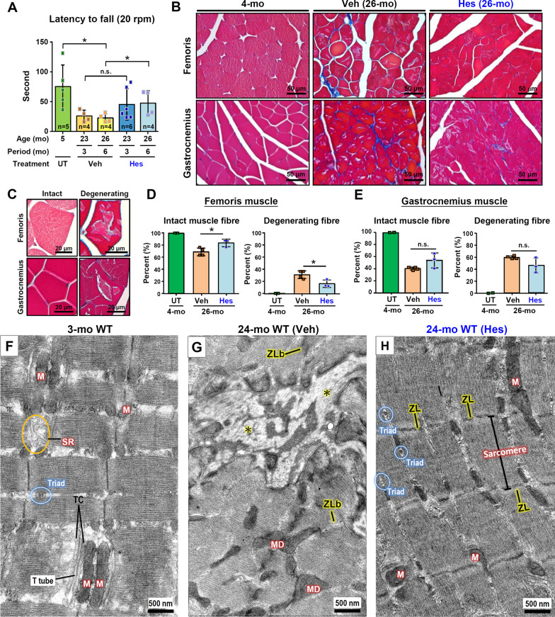
Fig. 4.
Hesperetin slows down skeletal muscle aging in old WT mice. A The Rotarod tests in old WT mice (n = 4–7 mice) were carried out after dietary hesperetin (100 mg/kg/day) or vehicle control food treatment for 3 and 6 months (started at 20-month old). B Masson’s trichrome staining of femoris and gastrocnemius muscles. The 21-month old WT mice were treated with dietary hesperetin for 5 months and sacrificed at 26-month old. C The representative micrographs showing muscle fibers with an intact and a degenerated morphology in the femoris and gastrocnemius muscles. D, E Quantification of intact and degenerating muscle fibers in femoris and gastrocnemius. Data are presented as mean ± SD. *p < 0.05; **p < 0.005 by one-way ANOVA with Bonferroni multiple comparison test in (D) and (E) or Student’s t test in (A). F–H TEM analysis of gastrocnemius muscle. F Young mice at 3-month old. G Veh-treated mice at 24-month old. Mitochondrial degeneration (MD) and fibrosis (*), which may be caused by myofibril degeneration and Z-line breakdown (ZLb), are evident and can be easily detected in the Veh-treated WT mice at 24-month old. H Hesperetin-treated mice at 24-month old. The age-related degeneration appears to be reversed as revealed by the presence of intact Z-lines (ZLs) and multiple normal-sized triads. M, mitochondria; SR, sarcoplasmic reticulum; TC, terminal cisternae of the SR. Scale bars, 500 nm

