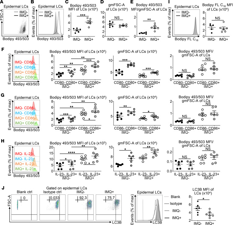
Figure 3. Elevated Langerhans cell lipid content in imiquimod-induced psoriasis-like skin.
Mice were treated as in Figure 2. (A–G) Mouse epidermal cells were stained with anti–MHC-II, anti–CD45.2, anti–CD80, and anti–CD86 Ab and Bodipy 493/503, which were analyzed by flow cytometry (n = 12, 3 independent experiments). Representative scatter plots (A), histogram (B), and MFI (C) of Bodipy 493/503 in epidermal LCs. The geometric mean FSC-A (gmFSC-A) (D) and the ratio of Bodipy 493/503 MFI to gmFSC-A (E) of LCs. Histogram, Bodipy 493/503 MFI, gmFSC-A, and the ratio of Bodipy 493/503 MFI to gmFSC-A of CD80–/CD80+ LCs (F) and CD86–/CD86+ LCs (G) from IMQ– and IMQ+ mice. (H) Epidermal cells were in vitro cultured with Golgi Stop for 4 hours, and they were stained with anti–MHC-II, anti–CD45.2, and anti–IL-23p19 Ab and Bodipy 493/503, which were analyzed by flow cytometry (n = 12, 3 independent experiments). Histogram, Bodipy 493/503 MFI, gmFSC-A, and the ratio of Bodipy 493/503 MFI to gmFSC-A of IL-23– and IL-23+ LCs from IMQ– and IMQ+ mice. (I) Epidermal LCs were incubated at 37°C with Bodipy FL C16 (1 μM) for 30 minutes, which were stained with anti–MHC-II and anti–CD45.2 Ab and analyzed by flow cytometry (n = 11, 3 independent experiments). Representative histogram and MFI of Bodipy FL C16 in epidermal LCs (dark gray filled, IMQ-untreated; black line, IMQ-treated). (J) Epidermal LCs were stained with anti–MHC-II and anti–CD45.2 Ab, which were further incubated with anti-LC3B Ab or rabbit IgG isotype control or none (blank control). Representative FACS analysis, histogram, and MFI of LC3B in epidermal LCs (light gray line, blank control; dotted black line, isotype control; dark gray filled, IMQ-untreated; black line, IMQ-treated; n = 12, 3 independent experiments). Two-tailed Student’s t test was performed. In (F–H), tests were considered significant with P < 0.05 after multiple testing adjustments by the FDR method. The data are presented as mean ± SEM. *P < 0.05, **P < 0.01, ***P < 0.001, ****P < 0.0001.

