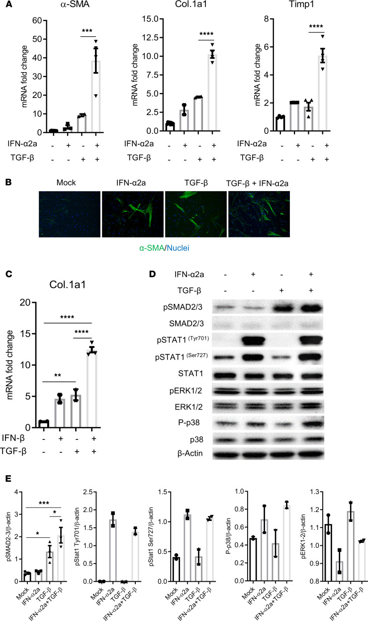
Figure 5. IFN-I and TGF-β synergistically activate primary human HepSCs.
(A and B) Rested HepSCs were treated with IFN-α2a (1000 U/mL) and/or TGF-β (1 ng/mL). (A) Expression of α-SMA, Col.1a1, and Timp1 was detected by RT-PCR. (B) Immunofluorescence of α-SMA (green) and nuclei (blue) in HepSCs exposed to TGF-β, IFN-α2a, and both (original magnification, ×20). (C) Rested HepSCs were treated with IFN-β (100 U/mL) and TGF-β (1 ng/mL). Expression of Col.1a1 was detected by RT-PCR. Data were normalized with Gapdh. (D and E) IFN-I increase TGF-β–induced activation of phosphorylation of SMAD2/3 in HepSCs treated with IFN-α2a and TGF-β. Rested HepSCs were treated with IFN-I and/or TGF-β for 60 minutes. (D and E) Immunoblot analysis (D) and quantification (E) of phosphorylation levels of SMAD2/3, STAT1 (Tyr701 and Ser727), p38 and ERK1/2, and of total SMAD2/3, STAT1, p38, ERK1/2, and β-actin in HepSCs. Histograms represent the average of 2–3 independent experiments; error bars indicate the SEM. Statistical analysis was performed with 1-way ANOVA and Fisher’s LSD test. *P < 0.05; **P < 0.005; ***P < 0.0005; ****P < 0.00005.

