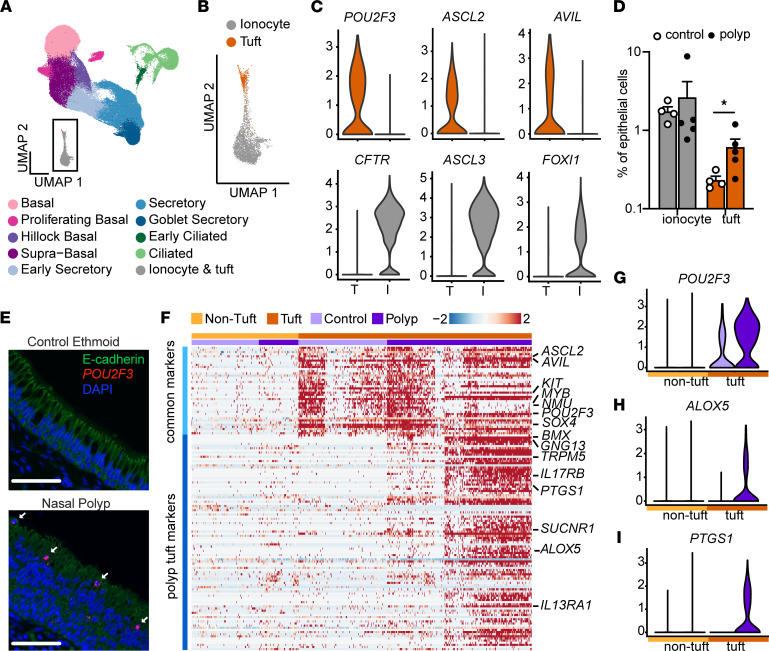
Figure 1. Single-cell sequencing reveals expansion and allergic activation of tuft cells in nasal polyps.
(A) Uniform manifold approximation and projection (UMAP) of scRNA-Seq of epithelial cells from healthy ethmoid sinus (control; n = 4) or nasal polyp (n = 5) (total cells = 116,358) reveals 10 cell types. (B) Expanded view of inset in A showing tuft cells and ionocytes identified by hierarchical subclustering. (C) Expression of tuft cell and ionocyte marker genes in tuft and ionocyte subclusters. (D) Percentage of ionocytes and tuft cells among total epithelial cells in control or polyp. Error bars indicate mean ± SEM. *P < 0.05 by Mann-Whitney t test with correction for multiple comparisons. (E) RNA in situ hybridization for POU2F3 (red) and immunofluorescence for E-cadherin (shown in green) identifies increased numbers of tuft cells (arrow) in nasal polyp epithelium as compared with control ethmoid (representative of 3 samples from 3 patients of each type). Scale bars: 60 μm. (F) Shared tuft cell marker genes (“common markers”) and differentially expressed genes (DEGs) (“polyp tuft markers”) in tuft (dark orange) and nontuft cells (light orange) from control (light purple) and polyp (dark purple) epithelium. Expression of representative (G) common tuft cell marker POU2F3 and polyp tuft markers (H) ALOX5 and (I) PTGS1.

