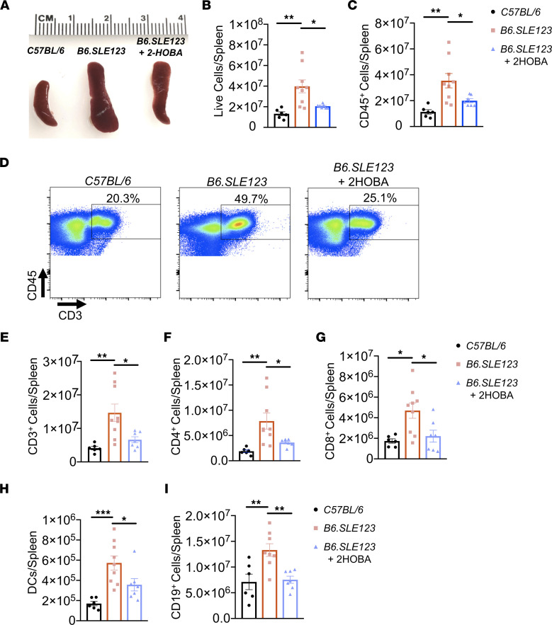
Figure 4. Scavenging of isoLG reduces splenic myeloid and lymphoid expansion in a mouse model of SLE.
Spleens and cells were isolated at the time of sacrifice from 32-week-old B6.SLE123 mice. Single-cell suspensions were prepared from freshly isolated mouse tissue via enzymatic digestion and mechanical dissociation. Live cell singlets were analyzed. (A) Representative spleens revealing a reduction in spleen size from B6.SLE123 mice treated with 2-HOBA. Quantitation of (B) live cells and (C) CD45+ cells. (D) Representative FACS plots for CD3+ T cells. Quantitation of (E) CD3+ T cells, (F) CD4+ T cells, (G) CD8+ T cells, (H) DCs, and (I) CD19+ B cells. Data were analyzed using 1-way ANOVA (n = 6–9, *P < 0.05, **P < 0.01, ***P < 0.001).

