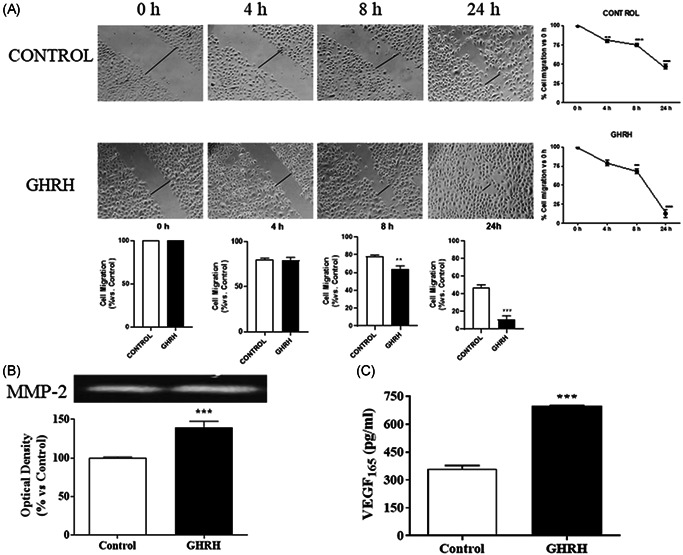
Figure 4.

Effect of GHRH on cell migration in RWPE‐1 cells. (A) Cells were damaged by mechanical scrapping and incubated in the presence or absence of 0.1 µM GHRH for the indicated times (4–24 h). Recovery of cell monolayer wounds was followed by microscopy. Representative images from four experiments are shown. (B) Cells were incubated with the peptide for 24 h. Total protein from cell secretion was subjected to zymography to detect the gelatinases. The expression of the latent isoform of metalloproteinase (MMP)‐2 was increased by 0.1 μM GHRH. A representative experiment is shown (n = 4). (C) Secretion of the vascular endothelial growth factor (VEGF) was evaluated by ELISA after incubation with GHRH (0.1 μM). The neuropeptide augmented the secreted VEGF165 by 90% at 24 h of the treatment. The diagrams represent the mean ± SEM of four experiments. **p < 0.01; ***p < 0.001 compared with control. ELISA, enzyme‐linked immunoassay; GHRH, growth hormone‐releasing hormone
