
Fig. 1 -.
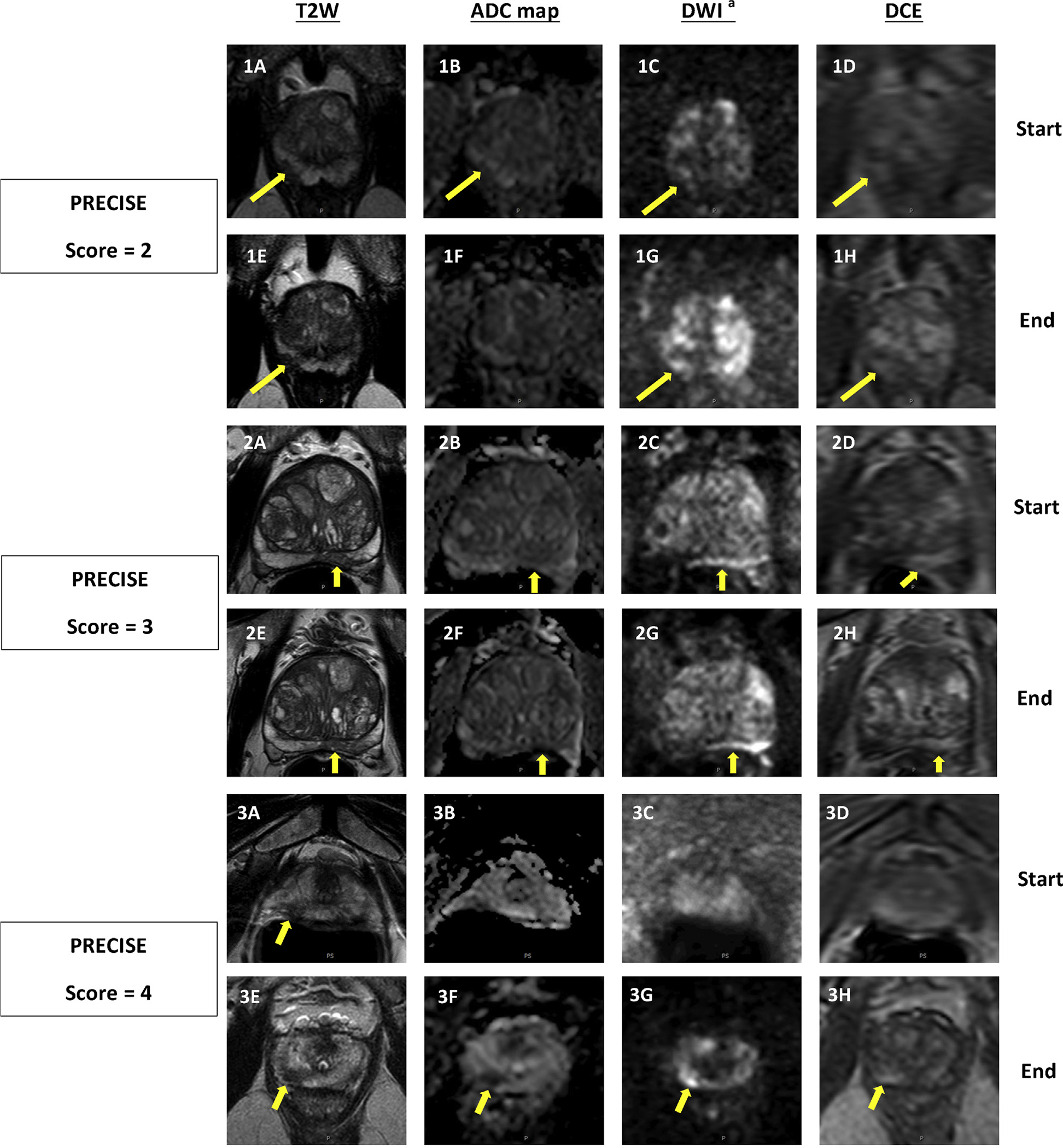
Three sets of intervals demonstrating how the PRECISE criteria are applied based on changing MRI features. Top row of each set (A–D) is at the beginning of the interval, bottom row (E–H) is corresponding to same pulse sequences at the end of the interval. (1) PI-RADS 3 lesion in right apical peripheral zone which was subsequently downgraded to PI-RADS 2 on follow-up MRI, as lesion is no longer visible on ADC map (PRECISE score = 2). PSA change (ng/mL): 5.23 → 9.00; PSAD change (ng/mL2): 0.11 → 0.15; MRI-targeted biopsy result: benign → benign. (2) PI-RADS 4 lesion in the left midbase peripheral zone remained stable on follow-up imaging (PRECISE score = 3). PSA change (ng/mL): 8.46 → 8.42; PSAD change (ng/mL2): 0.08 → 0.07; MRI-targeted biopsy result: benign → benign. (3) PI-RADS 4 lesion increased in size on T2W and became more prominent on DWI on follow-up MRI (PRECISE score = 4). PSA change (ng/mL): 5.16 → 5.81; PSAD change (ng/mL2): 0.15 → 0.14; MRI-targeted biopsy result: not targeted → GG1. ADC = apparent diffusion coefficient; DCE = dynamic contrast enhanced; DWI = diffusion-weighted imaging; GG = Gleason grade group; MRI = magnetic resonance imaging; PI-RADS = Prostate Imaging Reporting and Data System; PRECISE = Prostate Cancer Radiologic Estimation of Change in Sequential Evaluation; PSA = prostate-specific antigen; PSAD = prostate-specific antigen density; T2W = T2 weighted. a b = 1500s/mm2.