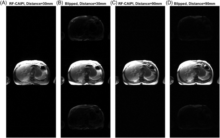
FIGURE 2.

RF‐CAIPI with GC‐LOLA (A,C) and blipped‐CAIPI (B,D) images acquired with a single band pulse (with MB3 blipped gradients for the blipped‐CAIPI acquisition and MB3 CAIPI phases and GC‐LOLA correction for the RF‐CAIPI acquisition). A,B, Acquired as for an SMS3 myocardial perfusion acquisition, with 30 mm inter‐slice distance between the three prescribed slices in the SMS slice group. C,D, Acquired with an inter‐slice distance of 90 mm. For the blipped CAIPI acquisition, there is significant leakage of the fat signal from the acquired slice into the top and bottom thirds of the image (corresponding to the locations of the other two simultaneously acquired slices of a MB3 acquisition) when an inter‐slice distance of 30 mm is applied. This is reduced when the inter‐slice distance is increased to 90 mm, which uses smaller blipped gradients
