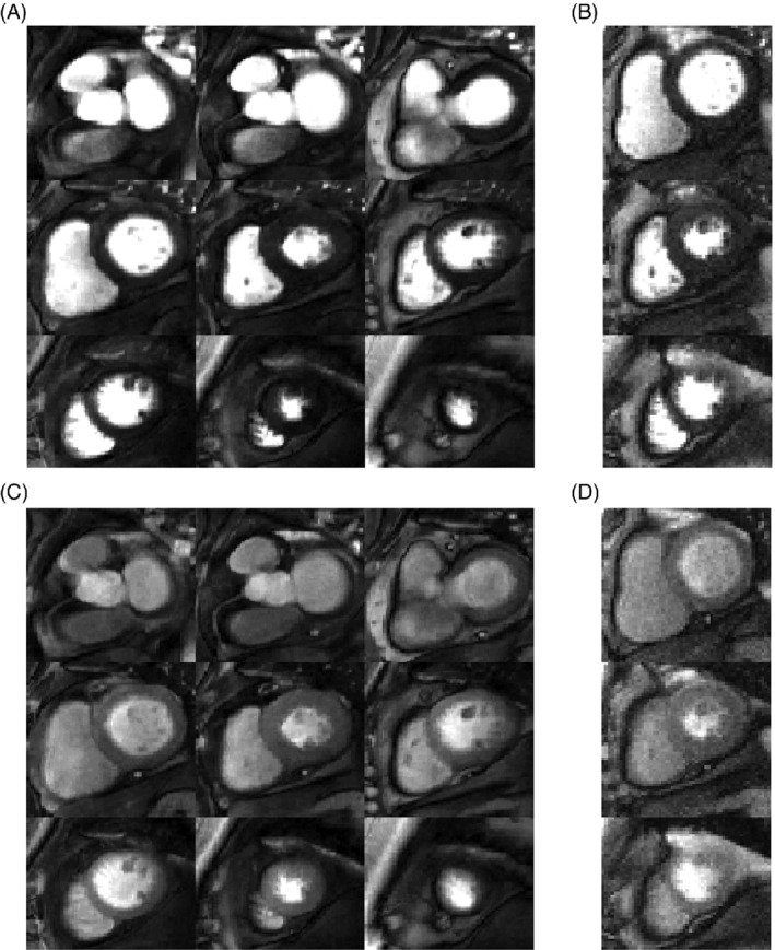
FIGURE 3.

Example perfusion images acquired in a patient under rest conditions, shown at peak enhancement of the left ventricular blood pool (A,B) and at peak myocardial enhancement (C,D). The nine‐slice high resolution (1.4 × 1.4 mm2) SMS sequence (A,C) was acquired first and the three‐slice conventional sequence with in‐plane resolution = 1.9 × 1.9 mm2 and GRAPPA reconstruction (B,D) was acquired after a 10‐min delay. For each dataset, the window width and level were kept constant for both timepoints. Image quality of all myocardial slices and both acquisitions were scored as 3.0 (= excellent image quality/no artifacts), except for the apical slice of the three‐slice acquisition, which was scored as 2.0 (= minor artifacts and of diagnostic quality), due to the partially obscured lateral wall in this slice
