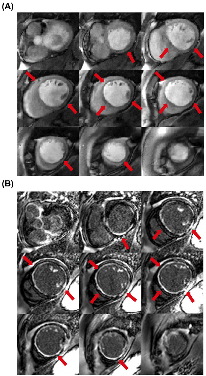
FIGURE 4.

A, SMS‐bSSFP perfusion images acquired under rest conditions in a patient with extensive subendocardial perfusion defects. B, Dark‐blood late gadolinium enhancement images 49 acquired in the same patient. Slice coverage is well‐matched between the two acquisitions. Regions of reduced myocardial signal enhancement indicated by red arrows on the perfusion images (A) match bright regions indicating scar on late gadolinium enhancement images (B)
