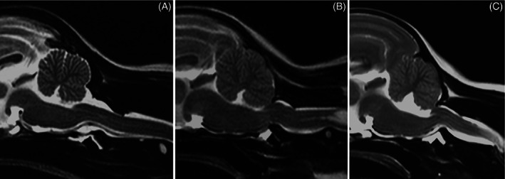
FIGURE 4.

Sagittal MR images (T2‐weighted TSE) of the craniocervical junction of a Maltese with a physiologic atlantoaxial joint ((A) control, VCI 0.11, CTR 0), of a Yorkshire Terrier classified as potentially unstable ((B) VCI 0.13, CTR 0.35), and of a Chihuahua diagnosed with AAI ((C) VCI 0.3). Note the loss of continuity of the cerebrospinal fluid dorsal to the dens, focal spinal cord compression, and medullary kinking as well as the marked cervical syringomyelia (B)
