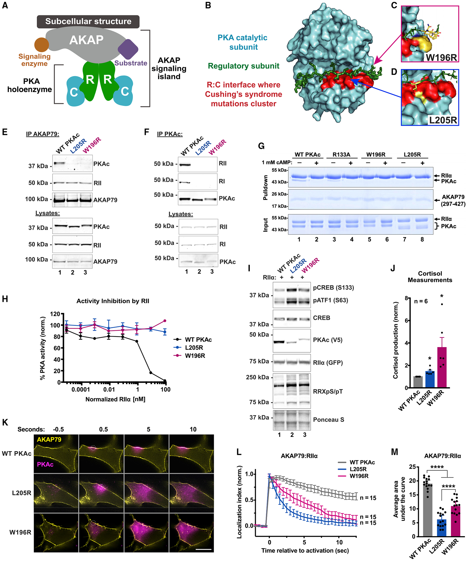
Figure 1. Cushing’s mutants are excluded from AKAP signaling islands.

(A) Model of AKAP signaling island. Regulatory subunits, R; catalytic subunits, C.
(B) Structure of PKAc bound to the inhibitory region of RIIβ (green). The W196R (C) and L205R (D) mutations fall within the red region.
(E) Immunoprecipitation of AKAP79 from H295R adrenal cells.
(F) Immunoprecipitation of PKAc from H295R cells.
(G) AKAP79 (297–427)-PKA complex formation in vitro ± cAMP.
(H) Changes in PKA-dependent phosphorylation of a fluorescently labeled peptide substrate upon increasing concentrations of RIIα. Mean ± SD; n = 4.
(I) Immunoblots of CRISPR PKAcα−/− U2OS cells expressing RII-GFP with either WT or mutant PKAc-V5. Mean ± 95% CI.
(J) Cortisol measurements from H295R cells infected with PKAc variants. Mean ± SE. *p ≤ 0.05, Student’s t tests after one-way ANOVA; n = 6.
(K) Photoactivation time courses in H295R cells. AKAP79-YFP, RII-iRFP, and PKAc tagged with photoactivatable mCherry were expressed. Scale bar, 10 μm.
(L) Quantitation of (K). Three experimental replicates.
(M) Integration of (L). Mean ± SE. ****p ≤ 0.0001, one-way ANOVA with Dunnett’s correction. See also Figure S1.
