Fig. 16.

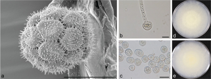
Morphology of Backusella luteola strain UoMAU6. a. SEM of sporangium; b. light microscope image of columella; c. light microscope image of sporangiospores; d, e. obverse and reverse of colony. — Scale bars = 20 μm.

Morphology of Backusella luteola strain UoMAU6. a. SEM of sporangium; b. light microscope image of columella; c. light microscope image of sporangiospores; d, e. obverse and reverse of colony. — Scale bars = 20 μm.