Figure 1.
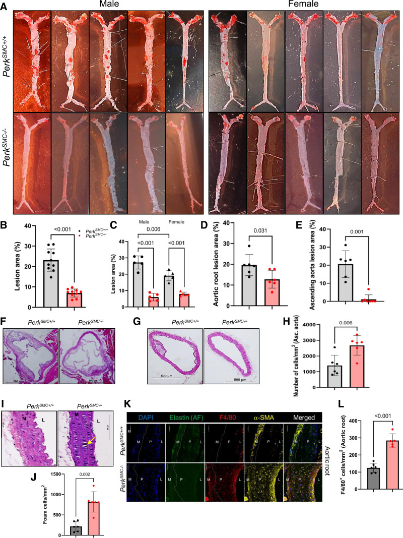
Smooth muscle cell (SMC)-specific deletion of Perk in hypercholesterolemic mice substantially reduces atherosclerotic plaque burden despite proatherogenic increases in serum lipid levels. A, En face Oil Red O staining of aortas shows significantly reduced plaque formation in PerkSMC−/− mice compared with PerkSMC+/+ mice, which is quantified in B (N=10, P<0.001, by unpaired Student t test with Welch correction). C, Oil Red O staining of aortas shows that Perk-deficiency prevents plaque formation to a higher degree in males than in females (N=5 per sex per genotype, analyzed by Kruskal-Wallis test followed by Dunn multiple comparisons test). H&E staining demonstrates that PerkSMC−/− mice have smaller atherosclerotic lesion areas in both the aortic roots (D, F, N=6, P=0.031, by unpaired Student t test with Welch correction) and the ascending aortas (E, G, N=6, P=0.001, by Mann-Whitney U test). H. Medial layers of PerkSMC−/− ascending aortas demonstrate higher cellular density that those of PerkSMC+/+ascending aortas (N=6, P=0.006, by unpaired Student t test with Welch correction). I and J, The medial layers of the ascending aortas of PerkSMC−/− mice contain a significantly higher number of foam cells compared with PerkSMC+/+mice (N=6, P=0.002, by Mann-Whitney U test). K and L, Immunostaining against the macrophage-specific marker F4/80 shows significantly higher staining in the aortic roots of PerkSMC−/− mice (P<0.001, using unpaired Student t test with Welch correction). Medial layers of the aortic roots and ascending aortas of PerkSMC−/− mice demonstrated more intense staining for α-SMA (smooth muscle α-actin) when compared with those of PerkSMC+/+ mice. Data are represented as mean±SD. AF indicates autofluorescence; L, lumen; M, medial layer; and P, plaque.

