Figure 2.
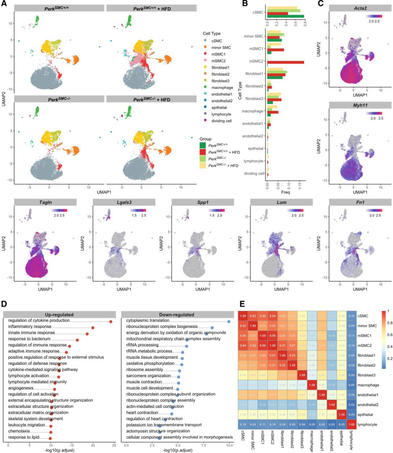
Single-cell transcriptomics of PerkSMC−/− and PerkSMC+/+ aortas at baseline and after hypercholesterolemia show loss of a specific subset of modulated smooth muscle cells (SMCs) in the PerkSMC−/− aortas. A, Modulated SMC clusters mSMC1 and mSMC2 expanded and appeared in the hypercholesterolemic PerkSMC+/+ mice with plaque formation, but mSMC2 did not appear in the PerkSMC−/− aortas with hypercholesterolemia. B, Perk deletion rescues the loss of contractile SMCs in PerkSMC+/+ aortas following HFD (top). mSMC1 cells are present at baseline in both genotypes and increase to a similar level following hypercholesterolemia. However, mSMC2 cells are rare at baseline in both mice but expand dramatically in PerkSMC+/+ aortas compared with PerkSMC−/− aortas. C, Expression patterns of contractile and SMC modulation markers in PerkSMC+/+ aortas following hypercholesterolemia. D, Top pathways in which DEGs of mSMC and cSMC clusters are enriched. E, Correlation plot indicates that mSMC1 and mSMC2 cells highly correlate with each other, as well as with contractile SMCs. Data are represented as mean±SD.

