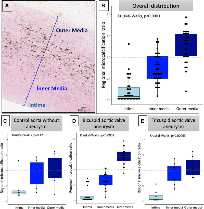
Figure 2.
Distribution of microcalcification in the proximal aortic wall of aneurysms. Assessment of the pattern of microcalcification across the aortic wall. A, Representative image of aortic wall stained with von Kossa, demonstrating significant microcalcification in the media, particularly the outer media, but not the intima. B, Across all samples assessed, as a ratio of total sample uptake, there was relatively little microcalcification of the intima, whereas there was significant deposition in the media, particularly the outer media. C through E, This pattern was consistent across aneurysm and control samples, with bicuspid aortic valve demonstrating a particularly strong preponderance for microcalcification in the outer media.

