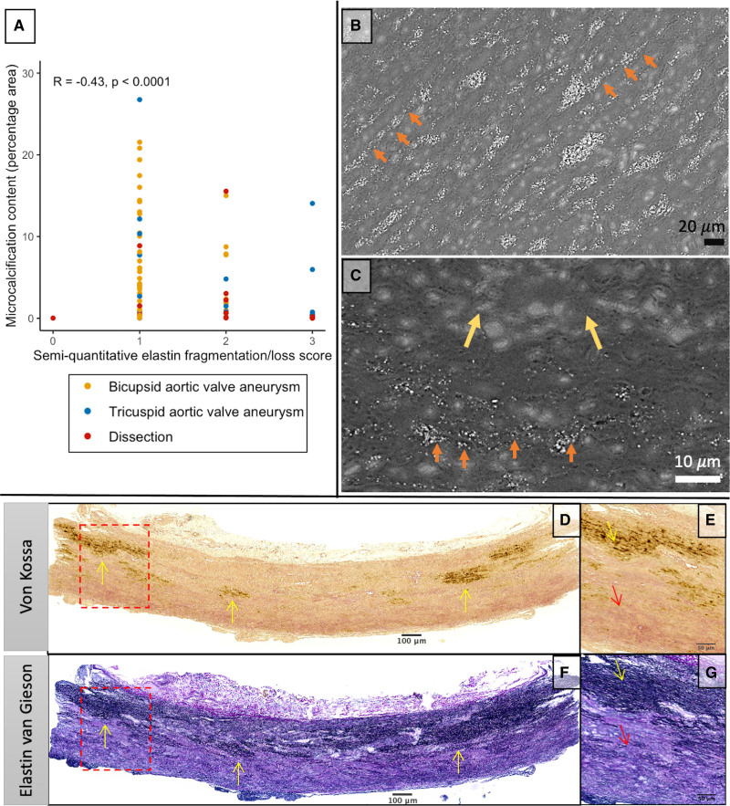
Figure 5.
Elastin fiber loss is associated with reduced microcalcification in severe aortopathy. A, Association between microcalcification content and elastin fragmentation/loss subcategory (Spearman ρ, −0.43; P<0.0001). B and C, Scanning electron microscopy from a sample with moderate histopathologic severity (B) with clear microcalcification precipitation on intact elastin fibers (orange arrows) and severe disease (C) demonstrating similar deposition along intact elastin but areas devoid of elastin with no microcalcification. D through G, Representative von Kossa (E and F) and elastin van Gieson (G and H) image from a patient with bicuspid aortic valve, severe elastin fiber loss, and microcalcification content of 0.4% percentage area (minimal). The microcalcification is clearly colocalized with areas of remaining intact elastin fibers (yellow arrows), whereas there is no microcalcification in areas devoid of elastin fibers (red arrows).

