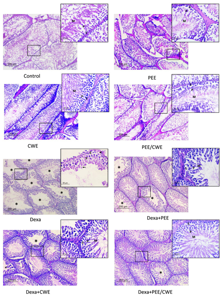
Figure 5.
H&E-stained histologic sections of seminiferous tubules in testicular tissues of the different studied groups. Control, PEE, CWE, or their combination showed mature sperms in all tubules (high spermatogenesis index). Higher magnification showed normal spermatogonia (S), multiple layers of spermatocytes (P), and mature sperms (M). The Dexa-treated model showed tubules with wide lumens lacking mature sperms (*). Higher magnification showed degenerated spermatogonia with focal cytoplasmic vacuolation (arrowhead), few spermatocytes (P), spermatids (arrow), and no sperms. Dexa + PEE and Dexa + CWE showed an improved spermatogenesis index and tubular lining in high magnification. The Dexa-treated group received combined products, and most tubules showed active spermatogenesis and intraluminal sperms. No degenerative changes were detected under high magnification. *: tubules with wide lumens and absent sperms; S: spermatogonia; P: spermatocytes; M: mature sperms; arrowhead: degenerative changes; arrow: spermatids. H&E; ×00, insert ×400).

