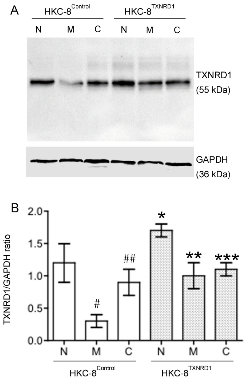
Figure 8.
Regulation of TXNRD1 protein expression by hypoxia and flavan-3-ol treatment in in both HKC-8TXNRD1 and HKC-8Control cells. Protein samples were prepared from both types of cells that were grown in normoxia or hypoxia in the absence (medium) or presence of 100 µM of catechin for 72 h. The levels of TXNRD1 protein and GAPDH were semi-quantitatively determined using Western blot analysis. (A) A typical blot of both TXNRDR1 and GAPDH protein of three separate experiments, (B) TXNRD1/GAPDH ratios in each group. N: Normoxia, M: Hypoxia with Medium, C: Hypoxia with Catechin. * p < 0.05 (HKC-8TXNRD1 vs. HKC-8Control cells in normoxia), ** p > 0.05 (HKC-8TXNRD1 in hypoxia vs. HKC-8Control cells in normoxia), *** p > 0.05 (Hypoxic HKC-8TXNRD1 with Catechin vs. Hypoxic HKC-8TXNRD1 with Medium) (One-way ANOVA, n = 3). # p = 0.0079 (HKC-8Control cells: Normoxia vs. Hypoxia/Medium), ## p = 0.0097 (HKC-8Control cells: Medium vs. Catechin) (two-tailed t-test).

