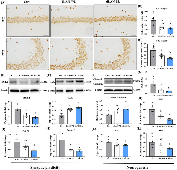
Figure 2.
Effect of dim light at night on hippocampal neurons in mice. (A) NeuN immunohistochemical staining in the CA1 and CA3 regions of the hippocampus in the different groups. Bar =50 μm. (B) The NeuN-positive cells in the hippocampal CA1 region (n = 9). (C) The NeuN-positive cells in the hippocampal CA3 region (n = 9). (D) Relative protein levels of BCL2 in the hippocampus (n = 3). (E) Relative protein levels of BAX in the hippocampus (n = 3). (F) Relative protein levels of cleaved-caspase3 in the hippocampus (n = 3). (G) The ratio of BCL-2 and BAX protein expression levels (n = 3). (H) The mRNA levels of Bdnf (n = 4). (I) The mRNA levels of Psd95 (n = 4). (J) The mRNA levels of Snap25 (n = 4). (K) The mRNA levels of Sirt1 (n = 4). (L) The mRNA levels of Dcx (n = 4). Differences were assessed using one-way ANOVA. The result represents the mean ± standard error of the mean. Values not sharing a common superscript letter (a,b) differ significantly at p < 0.05; those with the same letter (a,b) do not differ significantly (p ≥ 0.05). Ctrl: control group, dLAN-WL: dim white light at night group, dLAN-BL: dim blue light at night group.

