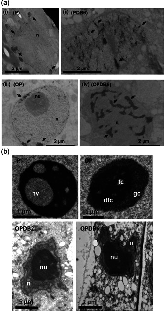
Figure 4.

Nuclear and nucleolar ultrastructure of primed and overprimed Medicago truncatula embryos. (a) Changes in chromatin distribution revealed by osmium ammine staining in the nucleus of M. truncatula embryos along the rehydration−dehydration cycle. In P and OP embryo axes, the nuclei of hydrated cells showed dispersed chromatin, with few heterochromatic domains at the nuclear periphery (i and iii, arrows). Chromatin condensation patterns were enhanced at the end of the dry‐back treatment (DB6) in both P and OP embryos (ii and iv, arrows). (b) Ultrastructural analysis reveals distinctive nucleolar architectures in imbibed P and OP seeds. For each sample, pools of 10 embryo axes were collected and 10−20 nuclei per sample were analysed. dfc, dense fibrillar centre; fc, fibrillar centre; gc, granular centre; n, nucleus; nu, nucleolus; nv, nucleolar vacuole
