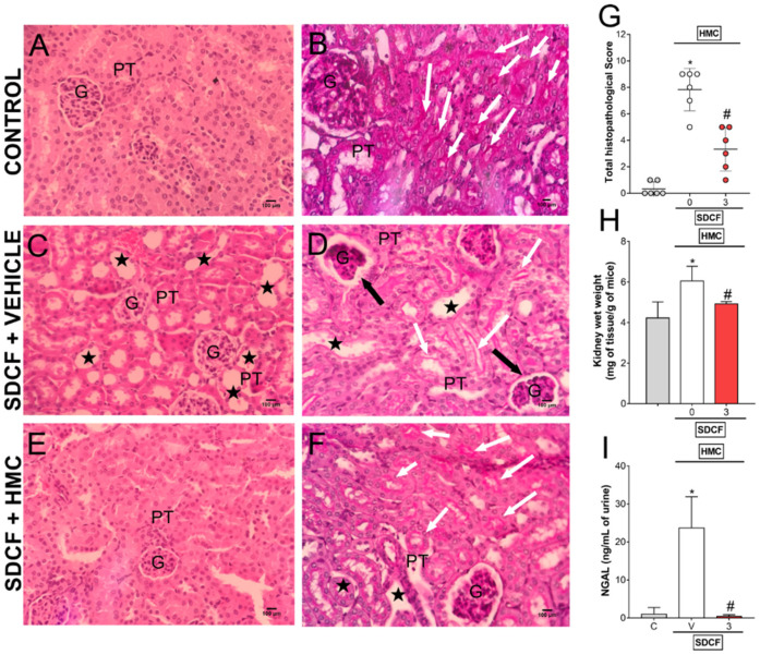
Figure 6.
HMC inhibits SDCF-induced renal histopathology, swelling and tubular cells cytotoxicity. Kidney samples were collected 24 h after the administration of SDCF for the evaluation of histopathology with H&E (A,C,E), and PAS (B,D,F) staining, total histopathological score (G), swelling (H), and NGAL urinary levels (I). Original magnification 40×; 100 µm scale. Stars show tubular dilatation; black arrows show glomeruli/Bowman’s capsule lesions; and white arrows show brush border differences in varied experimental groups. Data are shown as mean ± SD, n = 12 and n = 6 mice per group per experiment for histopathological analysis and swelling/NGAL, respectively, and are representative of two independent experiments. * p < 0.05 vs. control (C) group; # p < 0.05 vs. SDCF + vehicle (V) treated group; Kruskal-Wallis followed by Dunn’s post hoc test (G) and one ANOVA followed by Tukey’s post hoc test (H,I). G, glomerulus; PT, proximal tubule.

