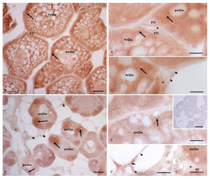
Figure 4.
Light micrographs of Anguilla anguilla ovaries from control (A–C) and exposed specimens (D–F). Immunolabeling with anti-17β-HSD antibody. (A–C) a wide and strong positive reaction was present in fully vitellogenic oocytes (fvOos) and early vitellogenic oocytes (evOos); a weak signal was evident in the follicular (FCs) and theca (*) cells as well as in the connective cells (arrowheads). (D–G) early vitellogenic oocytes (evOos) were immunolabeled, and the signal was in some areas of the cytoplasm as spots (arrow); meanwhile, weak positivity (arrow) was localized within previtellogenic oocytes (pvOos), follicular (FC), and connective cells (arrowheads). No signal was evident in the theca cells (*). (E) (insert) negative control: no labeling was evident in the ovary. Bars: (A) = 20 μm; (B,C,E–G) = 5 μm, (D) = 50 μm, (E) (inset) = 100 μm.

