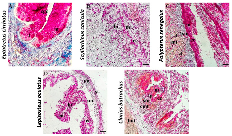
Figure 3.
Sections of intestine stained with Mallory Trichrome, 40×, scale bars 40 µm. (A) Mucosa (m), submucosa (sm), and Muscolaris serosa (ms) are the three layers of the gut of Eptatretus cirrhatus. The mucosa is arranged in zigzag ridges, with evident zimogenous cells (zgc), and scattered immune cells (l). (B) The intestine of Scyliorhinus canicula is separated into mucosa, submucosa, muscolaris, and serosa. The mucosa is organized in folds that are virtually obliquely oriented. A columnar epithelium (ce) of enterocytes forms the mucous layer (m). Scattered immune cells (l) are evident in lamina propria (lp). (C) The gut of Polypterus senegalus presents a mucosa (m) with columnar epithelium (ce), submucosa (sm), muscolaris (mt), and serosa (st). The mucous layer of the intestine shows goblet cells and columnar epithelial cells (ce). The thickness of the submucosa contains collagen fibers (cfs). (D) There are four strata in the gut of Lepisosteus oculatus: mucosa (m), submucosa (sm), muscolaris (st), and serosa (st). In the mucosa, longitudinal folds and a columnar epithelium (ce) accompanied by goblet cells can be seen. (E) The intestine of Clarias batrachus morphologically shows four laminae: mucosa (m), submucosa (sm), muscolaris (mt), and serosa (st). The mucosa of the intestine is folded and has a columnar epithelium (ce) comprising enterocytes and mucipar caliciform cells. Muscularis is organized in longitudinal (lmt) and circular (cmt).

