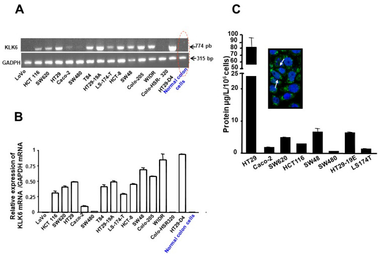
Figure 2.
Expression of KLK6 in human colon cancer cell lines. (A) Two micrograms of total RNA were reverse transcribed and PCR-amplified with KLK6 or GAPDH primers as described in Material and Methods. A major single PCR-amplified product of the predicted size (774 bp) for KLK6 was visualized after electrophoresis on a 2% agarose gel. GADPH was used as an internal control. Normal isolated epithelial cells do not express KLK 6 mRNA. Note that KLK 6 is present in SW620, a cell line from a lymph node of a primary adenocarcinoma from which SW480 was derived. (B) The amount of mRNA expression was quantified by densitometry of bands in comparison to the glyceraldehyde-3-phosphate dehydrogenase (GAPDH). Densitometry of mRNA bands were quantified from three independent PCR experiments presented as mean ± SEM. (C) Immunodetection of kallikrein-related peptidases 6 by colon cancer cell lines: Supernatants were collected from colon cancer cells in culture and KLK6 expression was estimated by sandwich-type ELISA (see Material and Methods). Protein values represent the mean concentration of KLK6 (µg/L) secreted by 106 cells, which were cultured for 24 h. Inset: shows confocal microscopic immunocytochemical localization of KLK6 in HT-29 cells (Magnification X630). Arrows show cytoplasmic, perinuclear staining of KLK6.

