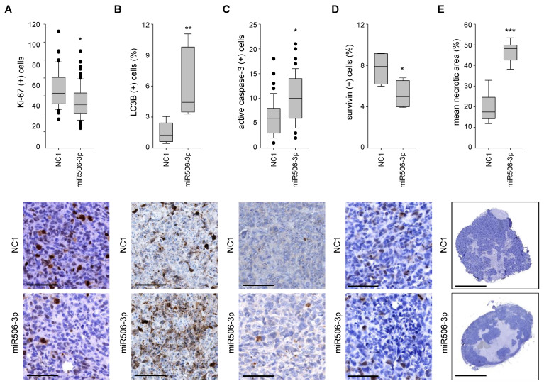
Figure 7.
Sections of explanted tumors, immunohistochemically stained with antibodies against (A) Ki-67, (B) LC3B (C) active caspase-3, and (D) survivin. (E) Necrotic areas (light blue) relative to the whole tumor. Upper bar graphs: results of overall quantitation; lower panels: representative microscopic pictures. * p < 0.05, ** p < 0.01 and *** p < 0.001. Scale bars: (A–D) 100 µm; (E) 2.5 mm.

