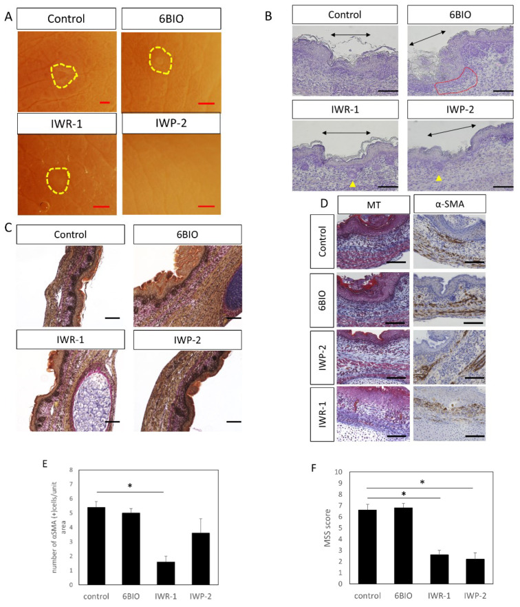
Figure 7.
E15 scar healing by administration of Wnt enhancer/inhibitor. (A) Observation by stereomicroscope. The scar is indicated by a yellow circle. Scars were not noticeable in the IWP-2 administration group. Scale bar = 1 mm. (B) Hematoxylin staining, scale bar = 100 μm. Black arrow: area of the scar. Yellow triangle: follicular structures observed within the scar. Red dotted line: area of thinning of the dermal structure in the scar. (C) EVG staining, scale bar = 100 μm. (D) Masson-Trichrome and α-SMA staining, scale bar = 100 μm. (E) Cell counting of α-SMA positive cells in wound. (F) Relationship between Wnt enhancer/inhibitor administration and wound MSS. * p < 0.05.

