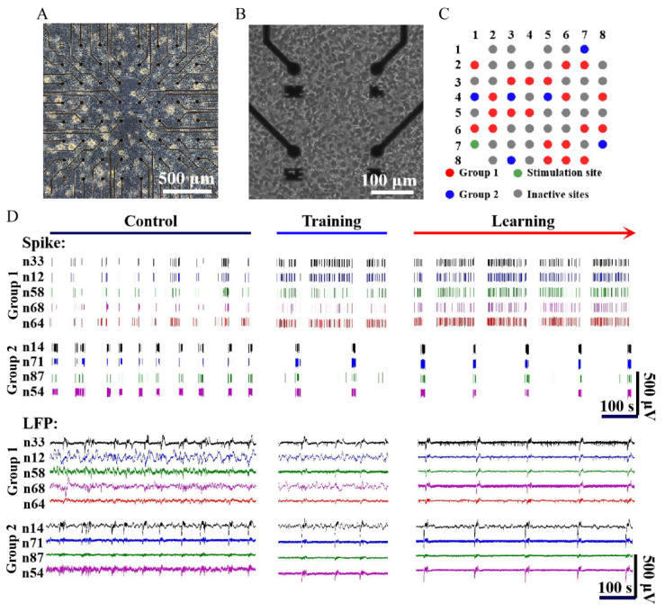
Figure 5.
Electrophysiology signals from the dissociated hippocampal culture before and after learning training. (A) Morphology of hippocampal neurons cultured for 1 week in vitro. (B) The neurons on the electrodes in (A) at higher magnifications. (C) The distribution and proportion of electrodes recording neuronal activities in Group 1 (red), electrodes recording neuronal activities in Group 2 (blue), inactive electrodes (gray), and stimulating electrodes (green). (D) Spikes and LFPs recorded by the neural sensor before and after training.

