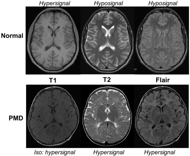
Figure 2.
MRI of a 5-year-old Pelizaeus–Merzbacher Disease (PMD) patient in comparison with an age-matched normal boy (Normal). In physiological conditions, the myelinated white matter has a hypersignal T1, hyposignal T2, and flair when compared to gray matter. In PMD, the hypomyelinated white matter has a normal hypersignal T1 appearance contrasting with an abnormal hypersignal T2 and flair. The corpus callosum is partially myelinated with a hyposignal T2 but not with flair.

