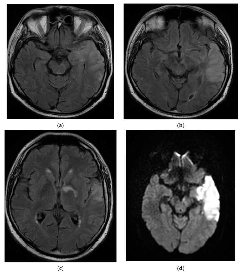
Figure 2.
MRI brain images with contrast showed aspects of T2 axial Flair hypersignal in the left temporal lobe (a,b), respectively, in the hippocampal and parahippocampal region (c). The lesion is hyperintense on the DWI axial image (d). MRI: magnetic resonance imaging; Flair: fluid attenuated inversion recovery; DWI: diffusion weighted imaging.

