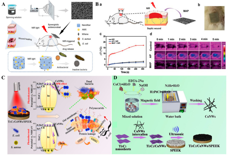
Figure 4.
(A) Schematic illustration of the fabrication of MAP nanofibrous membrane for antimicrobial therapy. (B) A model of the Balb/c mice infected with S. aureus (a); An image of the antibacterial dressing using the MAP nanofibrous membrane (b); NIR-induced temperature increase in the S. aureus-infected mice wounds (c), and the corresponding thermal images (d). Reprinted with permission from Ref. [91]. Copyright © 2021 Elsevier. (C) Schematic diagram for the NIR-activated antimicrobial mechanism of Ti3C2Tx/CoNWs/SPEEK. (D) Synthesis process for the coating of Ti3C2Tx/CoNWs heterojunction on porous SPEEK. Reprinted with permission from Ref. [92]. Copyright © 2021 Elsevier.

