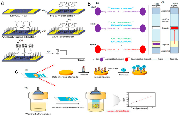
Figure 4.
(a) Real-time detection of rotavirus antibodies using a micropatterned reduced graphene-oxide-based field-effect transistor. a(i) Connection of PSE on MRGO surface, a(ii) DDT treatment, a(iii) immobilization of rotavirus-specific antibodies, a(iv) rotavirus capture, and a(v) real-time monitoring of rotavirus by electrical signaling. (b) Schematic illustration of the gold-aptamer-nanoparticle-based biosensor with (b(i)) colorimetric and (b(ii)) lateral flow response. (b(i(i))) Nanoparticles are functionalized with Sequence A (SH-5′ A12CCC AGG ACT AC T TTC 3′) and Sequence B (biotin -5′ GTG TTT CGG GAA G 3′ -SH) then aggregate by aggregating oligonucleotide (aptamer). (b(i(ii))) Oligonucleotide hybridizes with target nucleic acid (conserved enterovirus sequence) which (b(i(iii))) makes the nanoparticles, disaggregates, and changes their colors into red. (b(ii(i))) Aggregated nanoparticles are not able to move up through the lateral flow membranes, while (b(ii(ii))) in disaggregated form, they flow via lateral flow and bind to streptavidin due to biotin functionalization. (c) Norovirus detection by use of an impedance electrochemical biosensor. (c(i)) Immobilization of peptide self-assembly monolayers (SAMs) on the Au-working electrode. (c(ii)) Affinity strength of dropped norovirus, conjugated with the peptide on the gold screen-printed electrode (SPE), is measured using electrochemical impedance spectroscopy analysis. The figures were reprinted with permission from refs. [116,128,145], Copyright 2013, Elsevier; Copyright 20211, Multidisciplinary Digital Publishing Institute (MDPI); and Copyright 2018, Elsevier, respectively.

