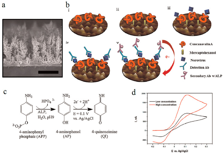
Figure 5.
Preparation and characterization of concanavalin A (ConA)-based electrochemical biosensor. (a) The scanning electron microscope (SEM) image. (b) Schematic illustration of biosensor: (b(i)) after fixation of ConA, (b(ii)) after blocking using mercaptoethanol (MCH), (b(iii)) after norovirus fixation, (b(iv)) after fixation of first Ab, and (b(v)) after fixation of secondary Ab using (c) ALP-labeled antibody transforms APP to AP, which is then oxidized and produces a current at the electrode that is identical to the quantity of NoV bound to the sensor surface. (d) Signal reading by use of cyclic voltammetry (CV). The figure was reprinted with permission from ref. [146], Copyright 2014, Elsevier.

