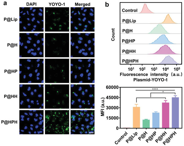
Figure 2.

Study on cellular internalization of plasmid delivery systems in cancer cells. a) CLSM images of HeLa cells treated by different plasmid delivery systems. CRISPR‐Cas9 plasmid was labeled by YOYO‐1 (green), and cell nuclei were stained by DAPI (blue). Scale bar: 36 µm. b) Flow cytometry analysis on HeLa cells treated by different plasmid delivery systems. HeLa cells were co‐incubated with plasmid delivery systems for 4 h. Untreated HeLa cells were served as a control. Data are mean ± s.d, n = 3. Statistical analysis was performed by using one‐way analysis of variance (ANOVA) with Tukey's multiple comparison test. *P< 0.05, ****P< 0.0001.
