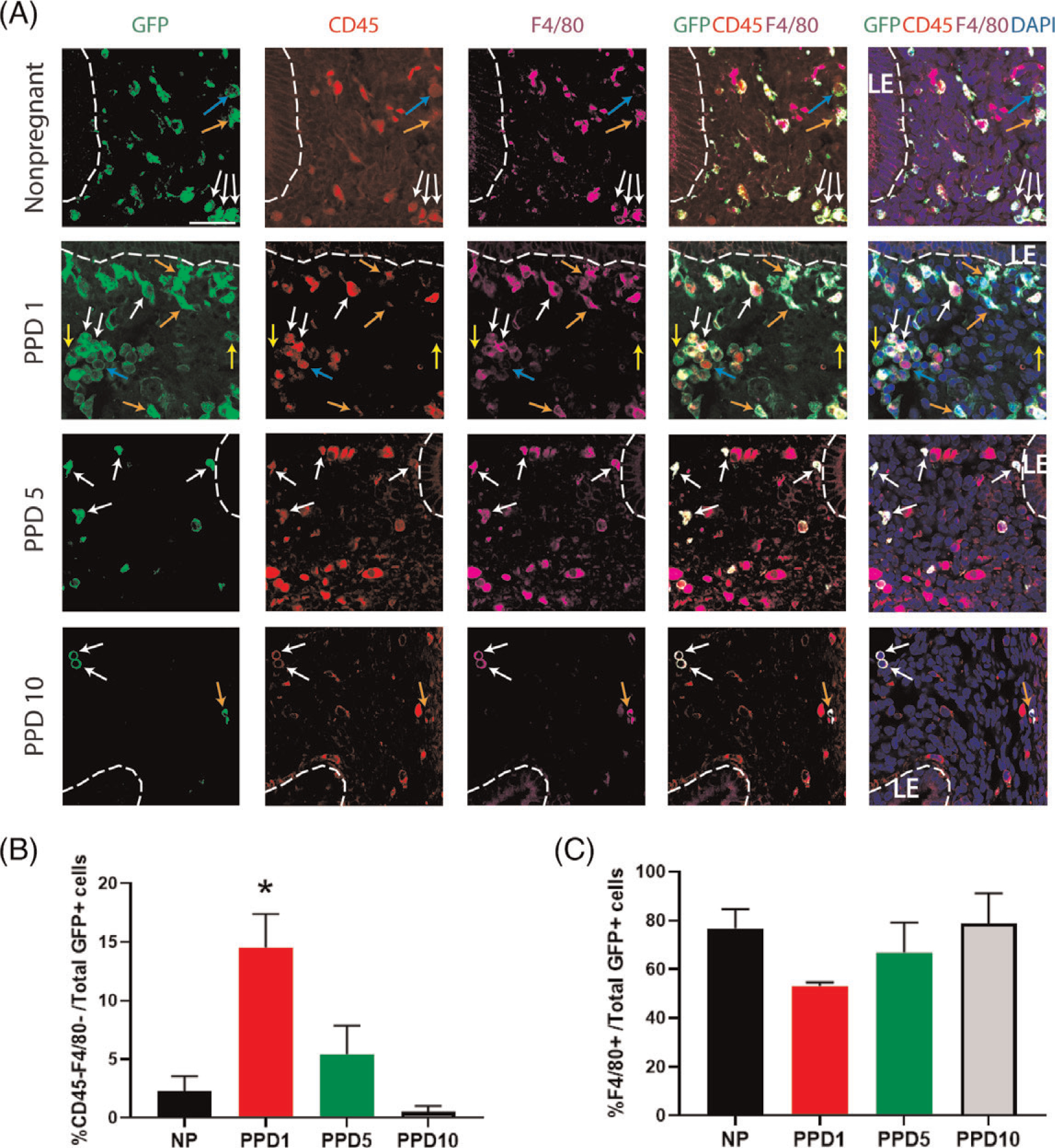
Figure 4. Co-immunolocalization of GFP+ BMDCs with CD45 and F4/80 markers.

(A) Immunofluorescence of uterine tissue sections showing colocalization of GFP-positive BMDCs (green), with CD45 pan leukocyte marker (red), and F4/80 macrophage marker (purple) across nonpregnant and postpartum day (PPD) 1, 5 and 10. Sections were counterstained with DAPI (blue) for nuclear staining. The dashed white line indicates the border of the luminal epithelium (LE). Blue arrows point to BMDCs (GFP-positive cells) that are positive for CD45 and negative for F4/80. Orange arrows point to cells that are GFP-positive, weakly positive for CD45, and F4/80-positive. White arrows point to cells that are GFP-positive, CD45-positive, and F4/80-positive. Yellow arrows point to nonhematopoietic cells that are GFP-positive but negative for either CD45 or F4/80 markers. (B) Quantitative summary of percentage of CD45-negative and F4/80-negative cells out of total GFP+ cells across different time points. (C) Quantitative summary of %F4/80+ cells out of total GFP+ cells. PPD1 (n = 5 animals); n = 4 in all other groups *P < 0.05 vs all other groups. Images were obtained with 40x lens. Size bar = 50 μm.
