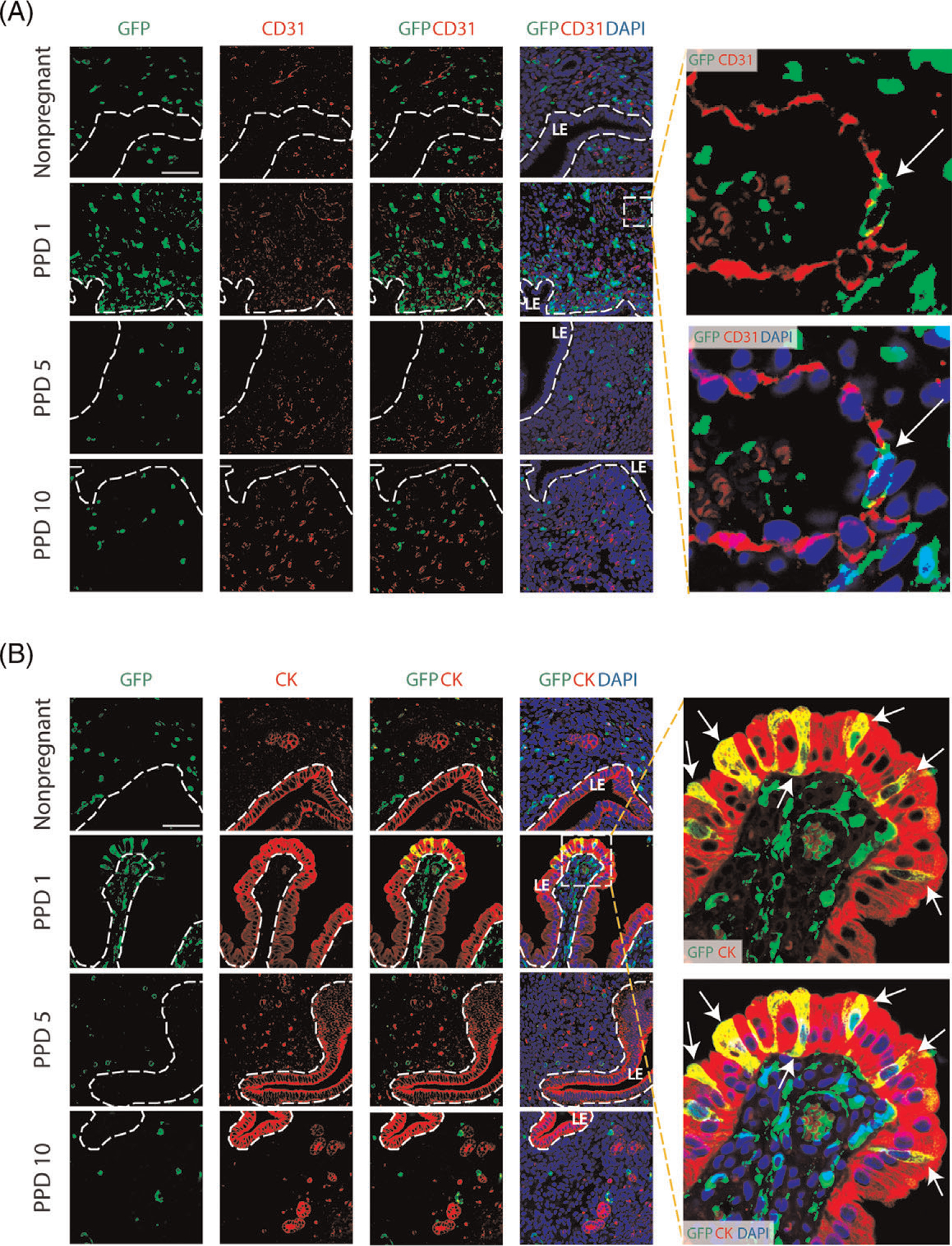
Figure 5. Co-immunolocalization of GFP+ BMDCs with CD31 and cytokeratin markers.

(A) Immunofluorescence of uterine tissue sections showing colocalization of GFP-positive BMDCs (green), with CD31 endothelial marker (red) across nonpregnant and postpartum day (PPD) 1, 5 and 10. Sections were counterstained with DAPI (blue) for nuclear staining. The dashed white line indicates the border of the luminal epithelium (LE). Images on the right are magnified images of the dashed square area. They show a bone-marrow derived endothelial cell integrated into a blood vessel on postpartum day 1 (white arrow), as visualized with GFP-positive cell colocalized with CD31 staining. (B) Immunofluorescence of uterine tissue sections showing colocalization of GFP-positive BMDCs (green) with the epithelial marker cytokeratin (CK) (red) across nonpregnant and postpartum day (PPD) 1, 5 and 10. Sections were counterstained with DAPI (blue) for nuclear staining. The dashed white line indicates the border of the luminal epithelium (LE). Magnified images to the right show GFP+ bone-marrow derived epithelial cells (white arrows) exhibiting typical epithelial morphology and integrated into the luminal epithelium on PPD1. Images were obtained with 40x lens. Size bar = 50 μm.
